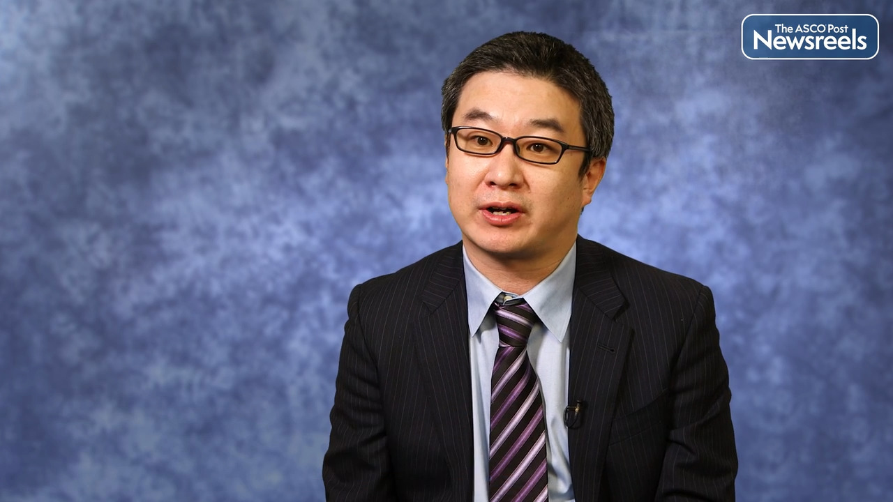
Jiye Liu, PhD, on Multiple Myeloma: Genome-Wide CRISPR-Cas9 Screening Identifies KDM6A as a Modulator of Daratumumab Sensitivity
2022 ASH Annual Meeting and Exposition
Jiye Liu, PhD, of Dana-Farber Cancer Institute, discusses study findings that demonstrate KDM6A regulates CD38 and CD48 expression in multiple myeloma. Dr. Liu’s team validated combination treatment with an FDA-approved EZH2 inhibitor plus daratumumab, which can overcome daratumumab resistance in preclinical multiple myeloma models, providing the rationale for combination clinical trials to improve patient outcome (Abstract 148).
Transcript
Disclaimer: This video transcript has not been proofread or edited and may contain errors.
We know that daratumumab is the first in class humanize the monoclonal antibody to target CD38 on myeloma cells. So the daratumumab triggers myeloma cell toxicity through the different molecular mechanisms including ADCC, ADCP and CDC, and the direct killing. And several clinical trials recently using the daratumumab alone and in combination with other agent to treat the newly diagnosed and the relapse, and the refractory myeloma cells then show the high efficacy. However, the relapse of the disease is commonly observed due to the daratumumab resistance. So we are seeking for the molecular mechanism of the daratumumab resistance in our project. We performed the genome-scale CRISPR screening in myeloma cells and find the genes named the KDM6A is most enriched in our daratumumab, positively select the gene list. And we found that the KDM6A is a histone 3 lysine 27 demethylase which activating the gene transcription.
Then we found that we knock out the KDM6A in the myeloma cells and can significantly downregulate the CD38 expression level on the myeloma cells. And associated with the increased H3K27me3 level on the CD38 promote area. We found that the KDM-60 knockout cells showed resistance to the DARAmediated ADCC in vitro and the in vivo mass model. Also by analyzing the [inaudible 00:01:55] in the KDM-60 knockout cells, we found that the KDM-60 also regulate CD48 expression, which is thought to be an NK-activating ligand in myeloma cells. So we knock out the CD48 in myeloma cells, attenuate the DARAmediated ADCC. So this data suggests that the KDM6A mediate ADCC not only by regulating CD38, but also modulate NK activity by the CD48 regulation. So how we can overcome the resistance induced by the KDM6A?
We know the KDM6A and EZH2 mediate the H3K27me3 level together in the cells and balance the genes transcription in the myeloma cells. So it is challenging to elevate the KDM-60 level in the myeloma patient cells. So we hypothesize that whether we can inhibit EZH2 to restore the CD38 or the CD48 expression level in the myeloma cells. So we used the one that FDA approved, the EZH2 inhibitor, to treat the KDM-60 knockout cells and find the CD38 and the CD48 expression level for restore also enhanced DARAmediated ADCC in these KDM-60 knockout cells. Our funding here identify a novel mechanism underlying the daratumumab sensitivity. This funding provided that the EZH2 inhibitor combined with the daratumumab to overcome the daratumumab resistance and for the translation to the clinical trial and improve the myeloma patient outcome.
The ASCO Post Staff
Stephen M. Ansell, MD, PhD, and Patrizia Mondello, MD, PhD, both of the Mayo Clinic, discuss the 20% of patients with follicular lymphoma (FL) who relapse early and experience a poor prognosis. The researchers found that FLs with high levels of IRF4 expression are associated with a suppressive tumor microenvironment, and selective IRF4 silencing restores antilymphoma T-cell immunity. Further investigation is warranted to identify the mechanisms by which IRF4 controls tumor immunity to develop precision therapies for this population (Abstract 70).
The ASCO Post Staff
Smita Bhatia, MD, MPH, of the Institute for Cancer Outcomes and Survivorship, University of Alabama at Birmingham, discusses study findings that showed key somatic mutations in the peripheral blood stem cell product increases the risk of developing therapy-related myeloid neoplasms (Abstract 119).
The ASCO Post Staff
Jia Ruan, MD, PhD, of Meyer Cancer Center, Weill Cornell Medicine, and NewYork-Presbyterian Hospital, discusses trial results demonstrating that the triple chemotherapy-free combination of acalabrutinib, lenalidomide, and rituximab is well tolerated, highly effective, and produces high rates of minimal residual disease (MRD)-negative complete response as an initial treatment for patients with mantle cell lymphoma, including those with TP53 mutations. Real-time MRD analysis may enable treatment de-escalation during maintenance to minimize toxicity, which warrants further evaluation. An expansion cohort of acalabrutinib/lenalidomide/obinutuzumab is being launched (Abstract 73).
The ASCO Post Staff
Tomohiro Aoki, MD, PhD, of the University of British Columbia and the Centre for Lymphoid Cancer at BC Cancer, discusses a novel prognostic model applicable to patients with relapsed or refractory classical Hodgkin lymphoma who were treated with autologous stem cell transplantation. The model has shown the interaction between the biomarker CXCR5 on HRS cells (Hodgkin and Reed/Sternberg cells, hallmarks of Hodgkin lymphoma) with specific follicular T helper cells and macrophages, a prominent crosstalk axis in relapsed disease. This insight opens new avenues to developing predictive biomarkers (Abstract 71).
The ASCO Post Staff
Eunice S. Wang, MD, of Roswell Park Comprehensive Cancer Center, discusses the outcomes of patients newly diagnosed with acute myeloid leukemia (AML) who were treated with cytarabine plus daunorubicin plus gemtuzumab ozogamicin (GO). These patients experienced higher rates of measurable residual disease–negative complete remission and complete remission with incomplete count recovery, compared to those treated with cytarabine plus idarubicin daunorubicin alone. Although adding GO was not associated with improved overall survival, longer follow-up is warranted to determine an absolute survival advantage of this regimen (Abstract 58).
