
Results from the first study using uEXPLORER to conduct total-body dynamic positron-emission tomography (PET) scans in patients with cancer suggested that it can be used to generate high-quality images of metastatic cancer. The research was presented at the Society of Nuclear Medicine and Molecular Imaging (SNMMI) 2020 Virtual Annual Meeting and published by Wang et al in The Journal of Nuclear Medicine.
Dynamic PET
While static PET provides a simple snapshot of radiopharmaceutical concentration, dynamic PET with tracer kinetic modeling can provide parametric images that show how tissue is actually behaving. Parametric images have the potential to better detect lesions and assess response to cancer therapy. This potential, however, has not been fully studied in the clinic because conventional PET scanners have a limited axial field of view and are not capable of simultaneous dynamic imaging of lesions that are widely separated in the body.
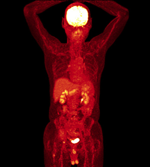
Photo credit: Getty
“The focus of our study was to test the capability of uEXPLORER for kinetic modeling and parametric imaging of cancer,” explained first study author Guobao Wang, PhD, Associate Professor and Paul Calabresi Clinical Oncology K12 Scholar in the Department of Radiology at the University of California, Davis. “Different kinetic parameters can be used in combination to understand the behavior of both tumor metastases and organs of interest, such as the spleen and bone marrow. Thus, both tumor response and therapy side effects can be assessed using the same scan.”
First Patient Results
A patient with metastatic renal cell carcinoma was injected with the radiotracer F-18 fluorodeoxyglucose (FDG) and scanned on the uEXPLORER total-body PET/CT scanner. The static PET standardized uptake value was calculated and kinetic modeling was performed for regional quantification in 16 regions of interest, including major organs and multiple metastases. The glucose influx rate was calculated and additional kinetic modeling was implemented to generate parametric images of the kinetic parameters. The kinetic data were then used to explore tumor detection and tumor characterization.
Multiple metastases were identified on the dynamic PET/CT scan, confirming that it is feasible to perform total-body kinetic modeling and parametric imaging of metastatic cancer. Parametric images of glucose influx rate showed improved tumor contrast over standardized uptake value in general, and specifically led to improved visibility of cancer lesions detection in the liver. Total-body kinetic quantification also provided multiparametric characterization of tumor metastases and organs of interest.
“Total-body dynamic imaging and kinetic modeling enabled by total-body PET have the potential to change nuclear medicine into a multiparametric imaging method, where many different aspects of tissue behavior can be assessed in the same clinical setting—much like the information gained from different sequences in a [magnetic resonance imaging] scan,” said study coauthor Ramsey D. Badawi, PhD, Professor in the Department of Radiology and Co-Director of the EXPLORER Molecular Imaging Center at the University of California, Davis. “The total-body parametric imaging technique is not limited to [F-18 FDG]; it is applicable to all radiotracers. It is also not limited to cancer but can be broadly applied to evaluate disease severity and organ interactions in many other systemic diseases. We expect a profound impact in the field of nuclear medicine and molecular imaging.”
Disclosure: For full disclosures of the study authors, visit jnm.snmjournals.org.