
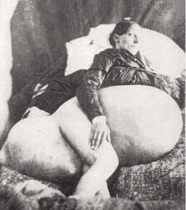
In June 1851, Philip J. Bruckner, MD, hired a daguerreotypist to photograph this 275-pound, 33-year-old woman, who had borne five children while developing this massive tumor. Dr. Bruckner learned of this patient when Charles Breech, MD, of Wellington, Ohio, presented her case at a medical meeting. Dr. Bruckner published the case history along with a woodcut illustration made from the photograph in the Ohio State Medical Society Transaction in 1851. It was one of the first uses of photography in a medical journal.
An earlier surgeon, Ephraim McDowell, MD, performed the first recorded ovariotomy and exploratory laparotomy in 1809, on a 47-year-old woman. Although the surgery was performed before the era of anesthesia and antisepsis, it was successful and the woman, Jane Todd Crawford of Green County, Kentucky, lived another 30 years.
Successfully performing ovariotomies defined a master surgeon in the early 19th century, and documenting large ovarian tumors became part of the culture of medicine during this era. ■
Excerpted from Oncology: Tumors & Treatment, A Photographic History, The Anesthesia Era 1845-1875 by Stanley B. Burns, MD, FACS.