
The text and photograph here are excerpted from a four-volume series of books titled Oncology: Tumors & Treatment, A Photographic History, The Antiseptic Era 1876–1900 by Stanley B. Burns, MD, FACS, and Elizabeth A. Burns. The photograph appears courtesy of Stanley B. Burns, MD, and The Burns Archive. To view additional photos from this series of books, visit burnsarchive.com.
During the 19th century, the most common occupational photograph of American physicians at work was the “dissection scene.” Photographs such as the one below were fashioned to hang in the physicians’ offices alongside their diplomas. From the 19th and throughout the first half of the 20th century, having a photographic portrait taken with one’s dissection cadaver was an initiation rite in medicine. The knowledge of anatomy separated a physician from a layperson, and these photographs provided evidence of the student’s entrance into the medical profession.
Human dissection has been interpreted for centuries in paintings and prints. Rembrandt’s The Anatomy Lesson and the prints of master anatomists of the 17th and 18th centuries serve as lasting symbols of serious medical study. With the advent of photography, medical students continued this custom, although the composition and posing changed with each decade. It was because of privacy concerns that this tradition ended.
It was not until the early 1880s that dissection itself became legal in most states. Laws permitting the practice were passed because of an outrageous grave-robbing incident that involved the stolen cadaver of former U.S. Senator John Scott Harrison. Mr. Harrison was the son of President William Henry Harrison (1841) and the father of Senator and future President Benjamin Harrison (1888).
Surgical Anatomy
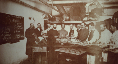
Although most dissection photographs depict medical students, the image shown here does not. This image portrays graduate physicians in an advanced course. These courses were given, much as they are today, to teach surgical anatomy. Knowledge of anatomy is the backbone of surgical excellence, and a surgeon must know what structures are under and to the sides of his knife. Anatomic dissection teaches the surgeon “standard” body proportions and to be aware that variances may occur. This photograph is quite remarkable and rare because of the information incorporated.
When looking at any vintage photograph, one should always question the “who, what, when, where, and why.” The inscription on the blackboard in the photo below provides instant answers to many of these questions. “N.Y. Post Graduate Hosp.” is written along the top. Underneath is the instructor’s name, “Prof. McAllister.” Following are two columns of student names, some of which are easily legible. The date is “May 1896,” and the final inscription, “Henry Curator Dead House,” supplies the last information. In the foreground is a table full of dissecting instruments.
Many dissection photographs in the 1890s were decorated with slogans, comic poses, and aprons emblazoned with states’ names, fraternity symbols, or funny comments. They were the remarks of anxious new students, who used humor to lessen the seriousness of their chosen profession.
During the final third of the 19th century, the New York Postgraduate Medical School was perhaps the best institution for advanced training in the United States, and many of New York’s leading surgeons and practitioners taught and practiced there. In the late 1890s, the establishment of the surgical residency training system at the Johns Hopkins Hospital brought surgical education into the 20th century.