Unlike ASCO’s Annual Meeting, symposia, and conferences, which highlight the current scientific advances in specific cancers and how they are improving cancer outcomes for the more than 18.1 million people worldwide diagnosed with cancer each year,1 ASCO Breakthrough: A Global Summit for Oncology Innovators examined how technology is transforming how cancers will be detected and treated in the near future.
ASCO’s first-ever international meeting, held in Bangkok, Thailand, on October 11–13, 2019, attracted nearly 500 attendees from more than 30 countries, working in a variety of disciplines, including information and computer science, artificial intelligence (AI), basic and translational research, social media, and biomedical engineering. More than 140 study abstracts were presented, many of which were conducted in the Asia-Pacific region. The 3-day meeting featured 13 sessions across such diverse topics as AI in Oncology and Therapeutics; The Future of Imaging in Oncology; and Microbiota, Immune Tonus, and Cancer, which were presented in conversational panel discussions to increase engagement with the audience. The next ASCO Breakthrough Summit will again be held in a city in Asia in 2021; more details to come.
“Breakthrough was extremely exciting and captured the intimacy of the early ASCO meetings, when you could fit all attendees in one room and everyone knew each other,” said Peter Paul Yu, MD, FACP, FASCO, Physician-in-Chief at Hartford HealthCare Cancer Institute, Past President of ASCO, and Chair of the Co-Host Committee for Breakthrough. “It didn’t matter whether you were a presenter or a member of the audience, everyone at Breakthrough was engaged in the topics being discussed. The speakers stayed around for all 3 days of the meeting, asking questions at the other sessions, and were available for spontaneous networking. It was very special.”
Attendees Most Interested in Sessions on AI
According to Dr. Yu, the sessions that drew the most audience interest were those focused on AI and machine learning. “At the end of the 3 days, we asked the audience what they were most interested in learning more about. We asked about bioengineering, nanotechnology, and precision medicine, but the topic attendees were most interested in hearing about was AI and machine learning and how that technology is going to transform many aspects of oncologic care and research and accelerate our ability to make progress in cancer care,” said Dr. Yu. “What I want to emphasize is that these AI systems are not meant to replace us. They are solutions to combine human cognitive ingenuity and creativity with the power of computers and data analytics. By efficiently performing routine tasks, they free highly trained oncologists to focus on the most complex problems and help to alleviate workforce shortages in low-income countries.”

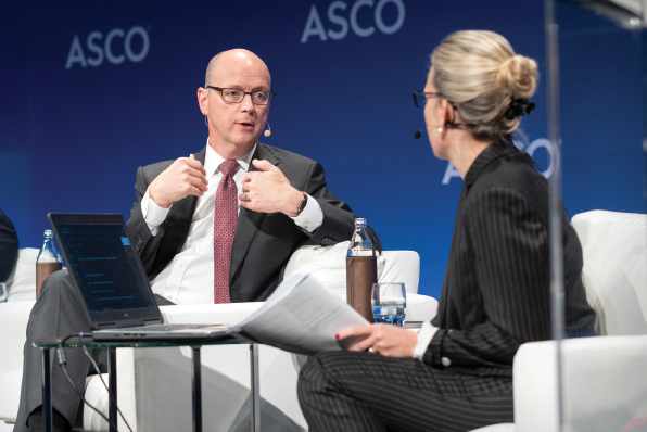
Gregory Sorensen, MD, explains the potential of machine learning in imaging in cancer care to moderator Rebecca A. Dent, MD, a medical oncologist at the National Cancer Center in Singapore, at the ASCO Breakthrough Global Summit. Photo by © Peerajit Photography.
The ASCO Post talked with three of the presenters at the Breakthrough meeting to learn more about the progress being made in their specific fields. Two of the presenters discussed how AI is transforming imaging in oncology: Gregory Sorensen, MD, Chief Executive Officer of DeepHealth, a Boston-based company focused on using machine learning to develop software products to assist radiologists, and Anant -Madabhushi, PhD, F. Alex Nason Professor II of Biomedical Engineering, Director of the Center for Computational Imaging & Personalized Diagnostics, and Professor, Departments of Radiation Oncology, Urology, Radiology, Pathology, General Medical Sciences, and Electrical Engineering at Case Western Reserve University. Another presenter, Siew C. Ng, PhD, Professor of Medicine and Therapeutics and Associate Director of the Center for Gut Microbiota Research at The Chinese University of Hong Kong, explained the role antibiotics play in disrupting the gut microbiome, which has the ability to control the immune system, lessening the effect of immune checkpoint inhibitors.
Dr. Sorensen: Role of AI in Oncologic Imaging
What is the potential of machine learning in imaging in cancer care?
It is nothing short of transformative. The breakthroughs in machine learning have been especially impactful in the imaging arena. We have had computer algorithms to analyze data in many forms over the past decade, but pattern recognition in images and image interpretation have traditionally been difficult problems for computers.
Starting around 2012, the first deep learning algorithms came into play, and that’s when things started to change. Now these image-recognition tools can, in some instances, be better than humans at recognizing certain patterns. This means we may finally have the opportunity for machines to learn to interpret images in a clinically useful way. And that’s important because imaging is central for so much in cancer care, from screening to follow-up, from surgical guidance to disease management. I think it’s the most impactful area that anyone could work on today in health care.

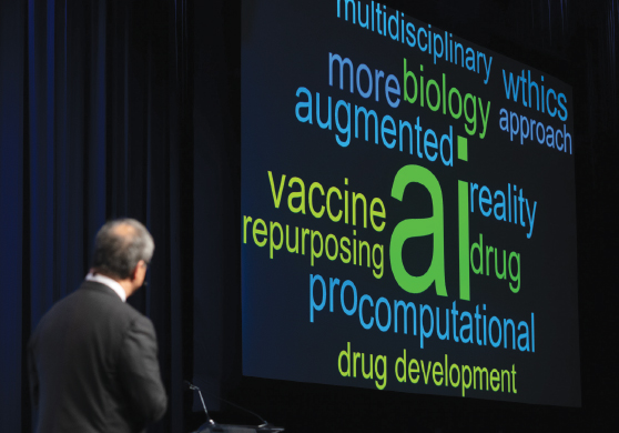
Peter Paul Yu, MD, FACP, FASCO, Chair of the Co-Host Committee for the Breaktrhough Meeting, on stage in Bangkok. Photo by © Peerajit Photography.
During your presentation at Breakthrough, you showed data from your study investigating the accuracy of human vs AI in interpreting mammography images for breast cancer. Your results found that the computer scored about 7 points higher on sensitivity and about 13 points higher in specificity.2 Does this technology have the potential to eliminate the need for radiologists in oncology care?
No, radiologists should not be worried about being replaced by a machine. There will always be the need for the human component in cancer care. Initial interpretation of screening images, which machine learning increasingly appears to be well suited for, is actually the part of the workflow that is the least interesting to my radiologist colleagues. They routinely report that the most rewarding and emotionally fulfilling part of their job is interacting with patients. Machine learning is just a tool to help clinicians take better care of patients and to help radiologists read films in a better way.
What are the most valuable opportunities for use of AI in oncologic imaging?
I do think breast cancer presents the greatest opportunity for this technology, because screening mammography has been so solidly proven to benefit women. Despite this benefit, the interpretation of x-ray mammography results can be uneven across different health-care settings, and it’s not desirable to have uneven performance in cancer care. So, if machine learning can help bring uniform excellence to that first step in diagnosis, it will have a big impact for many, many women. That’s why I picked breast cancer as the first focus area for our team, although it is hardly the only place in cancer care where machine learning can make an impact.

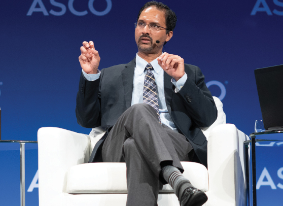
Among the crucial issues addressed by Anant Madabhushi, PhD, at the ASCO Breakthrough Global Summit was how artificial intelligence computational tools may help oncologists predict disease outcome and disease recurrence in their patients. Photo by © Peerajit Photography.
Other people at Breakthrough talked about machine learning in pathology, which is another area with the possibility of a lot of variability in interpretation. The beauty of machine learning analyses is that they can encapsulate all of the experience of many specialists—and that’s what we really love about AI.
You also talked about using AI algorithms to detect breast cancers a year before they are picked up on mammography. Please explain how these algorithms work.
Our hope is that, as machines get better and better at interpreting patterns and see more and more data, the algorithms may be able to see things that humans are uncertain about, overlook, or dismiss, perhaps because the tumor is so subtle in appearance it is confused with normal tissue.
We now have some evidence that this is feasible. In our study, we compared the accuracy of AI and radiologists in interpreting mammography findings; and, in fact, AI did do better than the human experts.2 If I were to use an analogy, one might think about trained observers trying to distinguish between, say, pictures of dogs vs pictures of wolves. By analogy, in our study, there were a number of findings in which the human interpreters thought they were seeing a dog and so didn’t need to worry about it. But the computer said, no, this finding is something to worry about, because this looks more like a wolf, and it turned out, in many cases, it was a wolf. The computer learned how to make the distinctions in patterns. This implies that AI could be further trained on the most dangerous cancers and eventually actually find them at a higher frequency. This is where we hope the field will continue to go, and these early results indicate the promise of this technology.
Dr. Madabhushi: The Future of Imaging in Oncology
Your presentation at Breakthrough centered on AI, computational pathology and radiology, and their implications in precision medicine. Please talk about the impact machine learning and computational pathology and radiology may have in precision medicine for cancer.
We are increasingly appreciating the amount of information embedded in routinely acquired data in the graphic images of computed tomography (CT) and magnetic resonance imaging (MRI) scans that allow us to use advanced, sophisticated, computational AI algorithms to tease out things the human eye cannot see. Over the past 5 years, there has been a particular interest in using AI approaches, not only to address questions about disease diagnosis, which is where a lot of the focus has been, but to start asking questions beyond that on how best to treat the disease, aggressively or nonaggressively.

Siew C. Ng, PhD (left), and ASCO President Howard A. ‘Skip’ Burris III, MD, FACP, FASCO, discuss how the use of antibiotics can disrupt the microbiome and negatively impact patients’ response to checkpoint inhibitors and other cancer therapeutics. Photo by © Peerajit Photography.
The focus of our work has been on developing and applying these computational tools to interrogate routinely acquired imaging data on CT and MRI scans, as well as on pathology slides. We want to go beyond diagnosis and ask specific questions about how we can predict disease outcome and disease recurrence and how we can identify patients who will have disease progression. These are critical questions because, from a clinician’s standpoint, the most important one is, how best to treat a specific patient: Do you give more aggressive therapy, do you give chemotherapy, or will the patient respond well to immunotherapy?
Anything we can do to help clinicians in this complicated decision-making process will offer huge value in oncology care. This is the essence of our work. We investigate how to use computer algorithms to interrogate the radiographic and pathologic images to tease out the features and backgrounds that the human eye may not be able to visually appreciate and to start to tell us about therapeutic response and benefit.
The development of these prognostic and predictive tools will allow us to realize the promise of precision medicine, which is to align the patient’s disease risk profile with the appropriate therapeutic management strategy.
How can computational radiology and pathology be used as predictive tools for treatment and outcome? When might these tools be ready for clinical use?
We recently published a study using AI technology with routinely acquired CT scans to identify patients with non–small cell lung cancer who were likely to respond to immune checkpoint inhibitors. We focused on three therapies: nivolumab, durvalumab, and pembrolizumab, and we were able to predict which patients were going to respond to them vs those patients who were not going to respond.3 However, more critically, we also found that not only could we predict response, but we were able to predict overall survival in these patients. In other words, the radiomic features we were mining from the CT scans were able to tell us not just about the short-term benefit of or response to immunotherapy, but we were able to prognosticate overall survival in these patients. This is of major value in oncology care, as just between 22% and 25% of patients currently respond to immunotherapy, and there is a huge overhead financial cost to these drugs. This is an area that is near and dear to us, and it is something we are spending a lot of time researching.
We investigate how to use computer algorithms to interrogate the radiographic and pathology images to tease out the features and backgrounds the human eye may not be able to visually appreciate.— ANANT MADABHUSHI, PhD
Tweet this quote
What will it take to move this technology to the clinic? I think we have to be able to demonstrate the validity of these algorithms across a spectrum of facts, multiple agents, and multiple patients, so validation is another critical piece of the puzzle. Also, physicians have to be convinced that this technology is not some flash in a pan and that it is not simply showing accuracy or performance metrics. We have to be able to link it back to what we know about the pathology and biology of disease.
It was a privilege to give a keynote lecture on “The Future of Imaging in Oncology” at the Breakthrough meeting. There was significant interest in AI technology, and I think it reflects the excitement in the clinical community for technologies like this. The current molecular diagnostic tests for cancers are tissue-destructive and expensive; you are sampling just a part of the tumor for gene-expression profiling, and it may not be the most aggressive component of the tumor. So, in the context of tumors with multiple subclones, the question is whether this molecular test really captures the entirety of the risk profile of a patient.
With the ability now of whole-slide imaging to scan routinely acquired hematoxylin and eosin (H&E)-stained pathology slides, we can digitally generate very high–resolution, high-density H&E slide images and apply AI algorithms to interrogate the slides in a way that has not been possible for pathologists before. We can start to capture the spatial architecture of the individual cells. We can look at texture patterns, heterogeneity patterns within the different tissue compartments within the stroma, the epithelium and individual nuclei. And what we are really able to do is convert this routine H&E slide into a digital quantitative biomarker, which represents the entirety of the morphologic diversity of the tumor across the entire landscape of the tumor.
However, before clinicians can become comfortable with the technology, they will want to understand the basis of its predictive ability. As we think about AI, it is important to establish the morphologic and molecular bases of these approaches. That is why a lot of our work is focused on looking at how the imaging relates to morphologic patterns on pathology and how the pathology relates to biologic pathways, genes, and proteomics. It is that multiscale assessment or association that is going to be very important as we think about the clinical adoption of these tools.
Dr. Ng: Microbiota, Immune Tonus, and Cancer
Your presentation at Breakthrough focused on how antibiotics can disrupt the microbiome and negatively impact patients’ response to checkpoint inhibitors and other cancer therapeutics. How should oncologists strike the proper balance between treating patients who may develop infections and concern that antibiotics may reduce the effectiveness of cancer therapies?
This is an important topic because antibiotics are commonly used around the world for various reasons. Patients with cancer may receive antibiotics to control an infection at some point during cancer therapy. It has been found that patients given antibiotics within 1 month before starting immune checkpoint inhibitors had a lower response to treatment. More important, use of antibiotics before immunotherapy was associated with worse overall survival in these patients.4
The challenge is for us to understand why this is the case. Although this remains unclear, it is likely that antibiotics cause prolonged disruption of the gut ecosystem and impair T-cell immune response against cancer, hence leading to less-favorable -outcomes.
It is important to further define the underlying mechanism. But, at least for now, clinicians planning to give immunotherapy to their patients should be cautious as to how and when to prescribe antibiotics, especially since antibiotics can cause rapid (within days) and profound, long-lasting (up to several months) changes to the composition of the microbial communities, affecting up to 30% of the bacterial species.
What should oncologists do if their patients need an antibiotic to treat an infection?
If the patient needs an antibiotic for a good indication, such as pneumonia or an abscess, then one has to treat the infection appropriately with antibiotics. On the other hand, if there is no strong indication, for example, the patient may have had a fever after receiving immunotherapy, the oncologist may be more vigilant about prescribing an antibiotic, especially prior to immunotherapy.
The impact of antibiotics on the gut microbiome is not limited to cancer therapeutics. Antibiotics are commonly prescribed over the counter in certain countries, and studies have shown that children given antibiotics early in life have a higher risk of developing obesity, inflammatory bowel disease, and metabolic diseases. It seems that the effects of antibiotics on the gut microbiome can persist from childhood to adulthood and may be associated with several chronic diseases.
During your Breakthrough session, the potential of using Fusobacterium nucleatum as a biomarker for the detection of cancer and as a predictor of treatment response was discussed. Please talk about this.
One emerging translational application of the gut microbiota is its use as a biomarker, and a good example is the use of Fusobacterium as a diagnostic biomarker for the early detection of colorectal cancer. Fusobacterium is associated with certain epigenetic phenotypes of colorectal cancer, including high degrees of microsatellite instability and CpG island methylation phenotype, which may provide an opportunity to develop diagnostic tools or treatment biomarkers for colorectal cancer. This marker has a high sensitivity for detection of not just colorectal cancer, but also precancerous lesions, including adenomas. Combining this marker with other existing tests, including the fecal immunochemical test, may lead to a high sensitivity. It is likely that the presence of Fusobacterium may serve as a diagnostic marker for colorectal cancer, but its role as a predictive biomarker for who might or might not respond to chemotherapy is unclear.
We need to incorporate this marker into clinical trials of patients with cancer treated with chemotherapy or immunotherapy. Then, we can determine whether this biomarker could help oncologists select the type of patients who may have a better response to a specific therapy.
Should patients’ microbiome be considered for eligibility into clinical trials?
If we can determine the response of patients to cancer therapy via their gut microbiome, we could spare some patients, such as those who are predicted to have an unfavorable response, for example, unnecessary treatment and/or related side effects and cost.
At the Breakthrough meeting, there was enthusiasm among oncologists surrounding the notion of collecting a patient’s stool sample before and during immunotherapy for different cancers. Our gut microbiota has close interactions with our immune system and cancer therapeutics. In the future, I believe this practice of collecting patients’ stool samples will be incorporated into clinical trials.5
This field will continue to grow, and gastroenterologists, microbiologists, bioinformaticians, scientists, and oncologists will be working closely together to bring microbiome medicine to the next level. ν
DISCLOSURE: Dr. Yu holds stock or other ownership interests in Apple, Contrafect, Google, IBM, Microsoft, and Oracle Corporation. Dr. Sorensen has been employed by DeepHealth; has held leadership roles for DFB Healthcare, Fusion Healthcare Staffing, IMRIS, Konica Minolta, and Siemens Healthineers; holds stock or other ownership interests in DeepHealth, DFB Healthcare, Fusion Healthcare Staffing, and IMRIS; has received honoraria from Reveal Pharmaceuticals; has served as a consultant or advisor to Hitachi Chemical; receives a share of patent royalties paid to Massachusetts General Hospital by General Electric; holds intellectual property interests in Imaging Biometrics and Olea Software; and has been reimbursed for travel, accommodations, or other expenses by Hitachi Chemical and Human Longevity. Dr. -Madabhushi has held a leadership role for Inspirata; holds stock or other ownership interests in Elucid Bioimaging and Inspirata; has received honoraria from AstraZeneca and Inspirata; has served as a consultant or advisor to AstraZeneca, Inspirata, and Merck; has received institutional research funding from Inspirata and Philips Healthcare; and holds institutional intellectual property interests licensed by Elucid Bioimaging and Inspirata. Dr. Ng reported no conflicts of interest.
REFERENCES
1. World Health Organization: Latest global cancer data: Cancer burden rises to 18.1 million new cases and 9.6 million cancer deaths in 2018. Available at www.who.int/cancer/PRGlobocanFinal.pdf. Accessed January 23, 2020.
2. Lotter W, Diab AR, Haslam B, et al: Robust breast cancer detection in mammography and digital breast tomosynthesis using annotation-efficient deep learning approach. arXiv:1912.11027, 2019.
3. Khorrami M, Prasanna P, Gupta A, et al: Changes in CT radiomic features associated with lymphocyte distribution predict overall survival and response to immunotherapy in non-small cell lung cancer. Cancer Immunol Res 8:108-119, 2020.
4. Pinato DJ, Howlett S, Ottaviani D, et al: Association of prior antibiotic treatment with survival and response to immune checkpoint inhibitor therapy in patients with cancer. JAMA Oncol 5:1774-1778, 2019.
5. Lynch SV, Ng SC, Shanahan F, et al: Translating the gut microbiome: Ready for the clinic? Nat Rev Gastroenterol Hepatol 16:656-661, 2019.

