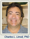
 The potentially devastating long-term consequences on cognitive function in patients with brain cancer following cranial irradiation led Charles L. Limoli, PhD, Professor of Radiation Oncology, University of California, Irvine, to study neural stem cell transplantation and how the procedure may prevent cognitive decline in this setting. Although an effective treatment for brain cancers, radiation therapy can sometimes induce debilitating side effects, including disruptions in memory and concentration and in the ability to carry out executive functions such as planning and multitasking, severely limiting quality of life.
The amount of cognitive destruction on still-developing brains is especially troubling. “Pediatric cancer patients, who have perhaps decades to live following successful treatment, can experience a loss of up to 3 IQ points per year,” says Dr. Limoli.
Since 2007, Dr. Limoli and his colleagues have been studying the effects of intrahippocampal transplantation of human neural stem cells in irradiated laboratory rats and then measuring their cognitive performance. The results of their study, published in the July 15, 2011, issue of Cancer Research,1 showed significant improvement in cognitive function in irradiated rats that received the neural stem cell transplants.
The ASCO Post talked with Dr. Limoli about his research.
Radiation and Cognitive Impairment
Explain how cranial irradiation damages cognitive function.
We use radiation on patients with brain cancer because outside of surgical resection, it’s really the only effective treatment that forestalls the growth of primary and metastatic lesions in the brain. Typically, radiation-induced late normal tissue injury in the brain, such as necrosis and edema, manifest at higher doses (> 40 Gy) and at protracted times (> 6 months) after radiotherapy is completed. However, at much lower doses (≤ 10 Gy) radiosensitive populations of neural stem cells are depleted, thereby limiting their capability to undergo neurogenesis, a naturally occurring process that contributes new brain cells to the hippocampus and helps to maintain cognitive health throughout life. Importantly, the depletion of these stem cells and the resulting cognitive deficits occur in the absence of any overt histologic or radiographically visible damage.
After survival, the next most important consideration for evaluating clinical outcome in cancer survivors may well be the quality-of-life issues related to cognitive function. The adverse effects of radiotherapy on the brain and cognition are progressive and difficult to reverse, and to date, there are no satisfactory long-term solutions for this serious clinical problem.
Study Specifics
How was your study conducted, and what were the results?
First, we selected a human neural stem cell (hNSC) that was derived from an embryonic stem cell and grew it to mass culture for transplantation. Then we irradiated athymic nude rats to induce cognitive deficits, and 2 days later, we stereotactically injected 100,000 stem cells into four sites in each hippocampus, for a total of eight injections in the brain (800,000 cells/animal).
At 1 and 4 months following transplantation, we subjected these animals to cognitive testing by giving them novel place recognition tasks to assess spatial recognition memory. Basically, the recognition task consisted of placing rats in a box containing two Lego blocks and allowing them to explore the location of these objects. After a period of familiarization, the rats were reintroduced into the same testing arena 5 minutes or 24 hours later, but after the position of the blocks had been moved to a different location. The capability of the rats to recognize and explore the object at the new location provides a very sensitive test for hippocampal memory.
What we found is that the animals that were given the human neural stem cells explored the new location of the block and the rats that were irradiated and not engrafted with hNSCs did not. The rats engrafted with hNSCs acted indistinguishably from unirradiated control rats.
When we sacrificed the rats to analyze their brains, we found that the hNSCs survived at significantly higher levels than we had anticipated. Unbiased stereology showed that 23% and 12% of the cells survived 1 and 4 months after transplantation, respectively. Immunohistochemistry was used to determine how these cells differentiated, and we found a significant number of new neurons and astroglial cell types. These data indicated that surviving engrafted cells turned into new neurons, new astrocytes, and new oligodendrocytes and that this combination of engrafted cells was able to restore cognition following irradiation.
We also found that while the stem cells migrated throughout the hippocampus, they tended to home to the very spot where normal neural stem cells exist, and migrated to the neurogenic niche within the brain. When we stained for activity regulated cytoskeleton-associated protein (Arc), which is a molecular determinant of memory, we found that our transplanted cells expressed Arc, suggesting that they had functionally integrated into the hippocampal circuitry. The presence of the Arc-positive cells suggested that the restoration of cognition was at least in part due to the functional replacement of cells lost or damaged as a consequence of irradiation.
In terms of how they were used in your study, are neural stem cells preferable to embryonic stem cells?
Our data does not indicate any significant differences between them, although there is a lower risk of teratoma formation with the human neural stem cells.
Next Steps
 Do you plan to study this procedure in human clinical trials?
Yes. I’m hoping to launch a phase I trial of this stem cell therapy in glioblastoma patients within the next 2 years. The protocol would be to transplant stem cells following brain tumor surgery and radiation treatments to assess safety and to restore cognitive function.
Are there applications for this procedure in other illnesses?
Absolutely. We recognized many years ago that this strategy might be an effective means to ameliorate “chemobrain,” a condition that is reported frequently in a growing population of cancer survivors subjected to a wide range of chemotherapeutic agents. These patients routinely exhibit a variety of cognitive impairments, believed, in part, to be caused by damage to the stem cells in the brain. We are actively engaged in determining the potential efficacy of using hippocampal stem cell transplants to reverse the adverse effects of chemobrain in this large cohort of patients.
Studies are also underway using hNSC transplantation to restore cognitive function following brain trauma, and evidence suggests the strategy may be beneficial in the treatment of Alzheimer’s disease and for alleviating side effects in any normal tissue subjected to radiation exposure.
While the field of regenerative medicine in radiation oncology is still very new, it holds a great deal of promise for the rehabilitation of many cancer survivors. Nevertheless, a significant amount of rigorous scientific work clearly remains to establish safety and efficacy in humans. ■
Disclosure: Dr. Limoli reported no potential conflicts of interest.
Reference
1. Acharya MM, Christie LA, Lan ML, et al: Human neural stem cell transplantation ameliorates radiation-induced cognitive dysfunction. Cancer Res 71:4834-4845, 2011.

