
When artificial intelligence (AI) is applied to medical images, it can improve physicians’ diagnostic capabilities as well as identify features that humans are unable to see. From identifying biomarkers to guiding treatment decisions, AI in medical imaging has already demonstrated numerous potential applications in research settings, but few of these tools have reached the clinic.
During a session at the Cleveland Clinic AI Summit for Healthcare Professionals, Benjamin H. Kann, MD, PhD, Associate Professor of Radiation Oncology at Harvard Medical School, explained some of the ways AI has transformed diagnostics and discussed its potential future impact on medical imaging.
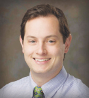
Benjamin H. Kann, MD, PhD
Dr. Kann, who also directs the Kann Lab in the Artificial Intelligence in Medicine (AIM) Program at Mass General Brigham and Harvard Medical School, described how clinicians can approach different types of AI applications in health care and assess how they may affect patient management.
Tiers of AI Applications
Dr. Kann outlined three tiers of AI applications for diagnostic purposes in health-care systems.
The first tier encompasses tools that identify features humans can readily see. These applications can often be deployed and monitored using established engineering and governance guidelines.
Dr. Kann explained that most tier one applications currently cleared by the U.S. Food and Drug Administration (FDA) for radiology are automation tools, with fewer cleared tools available for pathology.
In tier two, AI identifies features that humans have difficulty discerning on their own. As an example, he described a study of the real-world deployment of a fine-tuned pathology foundation model that could detect biomarkers in lung cancer based on histopathology alone.
Tier three represents AI applications that can identify features humans cannot see at all—because they involve predicting future events. These applications include decision-support tools and, according to Dr. Kann, have the highest potential actionability but also present the greatest challenges.
He noted that there are currently no FDA-cleared tier three applications in radiology. However, he pointed to the predictive ArteraAI assay as an example of a tier three application in oncology. The multimodal AI score can predict the benefit of adding hormonal therapy to radiotherapy for patients with localized prostate cancer and is based on large-scale evaluations using data pooled from five phase III trials.
Verifiability and Other Challenges With High-Tier Applications
In 2021, Dr. Kann and colleagues described an AI translational gap defined by shortcomings in validity, usability, and utility. Five years later, he said the field has underestimated a fourth factor: verifiability. “What also really matters is how verifiable these tools are,” he said. “The more AI changes what we do, and by that I mean the decisions we’re making, the less able we are to check it.”
He explained that tier one applications can be readily verified on a case-by-case basis, but verifiability decreases with the higher tiers. Tier two applications can be assessed only in aggregate through local validation. Although such assessment is feasible in real time, it is resource-intensive. Tier three applications, on the other hand, may be auditable only months or years later, when it becomes possible to determine whether the AI performed as intended.
This difference in verifiability contributes to the translational gap and may help explain the greater adoption of tier one models in health care.
Additionally, tier three applications are often subject to data set drift and shift, in which changes in the patient population, demographics, or clinical practice alter the underlying data and raise questions about whether the original training data remain applicable. As a result, the performance of prognostic models may degrade over time. Dr. Kann stressed that once models are deployed, ongoing monitoring of the AI is necessary.
He also explained that as the biologic understanding of disease increases, statistical and training stability may decrease. For example, once a new disease subtype is identified, a model that has not undergone additional training may be unable to accurately predict how that subtype will respond.
“As we learn more and more, this becomes statistically a much harder problem to solve, so this gives me a lot of skepticism about some of these prognostic and predictive approaches,” Dr. Kann commented.
Foundation models, on the other hand, can reduce the need for labeled data through fine-tuning, but they do not address other challenges, including event scarcity, confounding, data set drift, follow-up time, and signal ceilings.
With multimodal AI models, incorporating multiple types of data can raise the signal ceiling but also requires more data to reach that ceiling. This introduces greater potential for confounding, redundancy, missing data, estimation variance, and measurement noise across the different platforms used to collect or extract data.
Opportunities With Tier One Applications
Dr. Kann noted that tier one applications can be particularly useful when linked to actionable findings. These tools can also uncover features that were not previously routinely measured. For example, Dr. Kann’s team used a segmentation algorithm to measure the temporalis muscle on routine brain MRI scans from pediatric patients with brain tumors, using the measurement to assess body composition for prognostication. They found that 73% of survivors had sarcopenia at one or more time points, and 84% had a normal or overweight body mass index at the time. Sarcopenia was associated with endocrine disorders, poor physical functioning, and mortality. The group is now planning a prospective trial (STRIVE-AI) to use temporalis muscle measurements to identify patients who may benefit from exercise and nutritional interventions.
Dr. Kann’s colleagues applied the same concept to the thymus, which is visible on chest CT scans routinely obtained for patients with lung cancer. An AI-derived score of thymic health was significantly associated with immunotherapy response, and trials are being designed to evaluate its use in treatment decision-making.
What a Clinician Needs to Know About an AI Model Before Acting
Dr. Kann described an example involving a model used in his own lab to detect extranodal extension on CT scans of patients with head and neck cancer. In one case, the model incorrectly indicated that extranodal extension was present.
Before a model is deployed, Dr. Kann stressed, clinicians need access to several types of information to appropriately interpret its output and use it in patient management. These include the model’s raw output data; calibration information to indicate the certainty of its predictions; conformal prediction, which allows the model to abstain when presented with unfamiliar cases; and information about the training data to help clinicians recognize the model’s limitations.
Clinicians should also know whether a model fails in predictable ways. “If you know where your model is likely to fail, you can build out the specification of the model for the user,” he said. “[Unpredictable failures are] more concerning, because then you don’t really know when it’s failing and when it’s not.”
Dr. Kann added that clinicians should understand the potential risks associated with applying an AI algorithm. “Nobody would be comfortable with an AI guiding where to make an incision if it was only correct 80% of the time,” he explained.
The tier system, he said, provides a framework for determining whether and when a model can be used in clinical practice. “Knowing which tier you’re evaluating will drive the questions you should ask and the resources you need for safe and effective clinical use,” Dr. Kann explained.
He reiterated that tier one models can be used safely with appropriate monitoring and governance guardrails, provided that health systems budget for ongoing monitoring as well as licensing costs. For tier two applications, health-care professionals should validate the model in their local population and account for uncertainty. “A model that admits uncertainty beats one that’s confidently wrong,” he said. For tier three applications, he recommended focusing first on cohort development before building the model and requiring both prognostic and predictive clinical evidence, ideally derived from randomized trials.
“The future of cancer AI diagnostics depends less on whether models can predict—and more on whether clinicians and health-care systems can use predictions responsibly,” Dr. Kann concluded. “We have a lot of models coming out. The power is there, the capability is there, but we really need to think about whether health-care clinicians and systems can use these outputs [effectively].”
